Case Study # 3
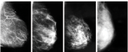
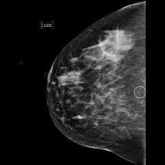 Mammogram
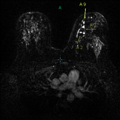 MRI
MRIThis is a 39-year-old woman with family history of breast cancer in her sister at age 40. Her mammogram reveals dense breast tissue, with no suspicious findings:


She is at increased risk for breast cancer because of her family history. Her breast tissue is dense, which limits the accuracy of her mammogram. The radiologist reading her mammogram recommended that she have an additional screening test, breast MRI, for high-risk evaluation:

There is a suspicious region in the upper inner left breast on this MRI image (4 bright white spots between the arrows). She underwent a needle biopsy with MRI guidance. Diagnosis: DCIS (ductal carcinoma in situ) with microinvasion– i.e. early breast cancer. Even knowing where the cancer is, it could not be seen on the patient’s mammogram.