Case Study #2
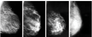
 Mammogram
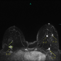 MRI
MRIThese are mammogram images from a 63-year-old woman with no family history of breast cancer. She has a screening mammogram every year, and believes that if she were to develop breast cancer, it would be found early since she is so diligent about her check-ups.
Though she is post-menopausal, she still has dense breasts (as do 1/3 of women over 50 yrs old). She was never told that she has dense breasts, and she did not realize that a mammogram on dense breasts will not detect up to half of all breast cancers. She had no idea that having a yearly screening breast ultrasound or MRI in addition to a yearly mammogram would give a much higher likelihood of finding an early cancer in her breasts.
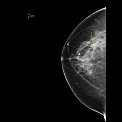
On this screening mammogram, the radiologist spotted a very subtle area of “architectural distortion” in her left breast. The arrows point to this distorted area within the dense white breast tissue:


She had a breast ultrasound, which found a mass in this area of the left breast, and she had a needle biopsy of the mass. The diagnosis was invasive ductal carcinoma (breast cancer). She had an MRI before her surgery, to evaluate the extent of the cancer:

Arrow A13 points to the biopsied cancer in the left breast. Arrow A17 points to a second mass in the left breast that was not visible on her mammogram. Ultrasound was performed, which found this second mass, and a needle biopsy diagnosed an additional cancer.
Arrow A20 points to a tiny spot in the right breast which was biopsied with a needle under MRI guidance. This was benign (not cancer).
Arrows A15 and A19 point to abnormal lymph nodes. These were biopsied, and contained metastatic cancer.
This patient was treated with a left mastectomy, radiation to the axillary lymph nodes, and chemotherapy. Although the patient was diligent about having her yearly mammogram, her cancer was not detected early, and her chances of long-term survival are now lower.