Case Study # 4
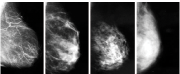
 Mammogram
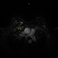 MRI
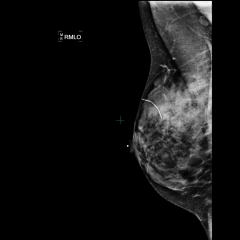
MRIThese are screening mammogram images from a 48-year-old woman with family history of breast cancer in her mother. She had a benign lump removed from her right breast several years before. The scar is designated by a sticker which shows up on the films below (very dense breast tissue, but negative mammogram) as a curved white line:


The radiologist reading her mammogram knew that this mammogram had a high likelihood of missing a cancer, due to the dense white breast tissue which could mask a tumor. In addition, the patient was at high risk due to the family history of breast cancer in her mother. The radiologist recommended that the patient have an MRI of her breasts, for high-risk screening. Her gynecologist agreed, and after battling with her insurance company to cover it, the patient had the MRI a few months later:

After looking at the above MRI image, the radiologist was unhappy. Look at all of the small white dots lighting up in the patient’s breasts! How can one possibly decide if one of them is cancer? The radiologist wished he’d never recommended the MRI.
But then……

…..one bright mass in the right breast (yellow arrow) seemed to stand out from the others. The radiologist called the patient back into the office to have a targeted breast sonogram in that particular area of the inner right breast. Lo and behold, the sonogram found a 9mm mass (yellow arrow):

A needle biopsy was performed, and the mass was malignant (invasive ductal carcinoma). At surgery, there was no spread to the lymph nodes under the arm. Final result: Stage I breast cancer, with an excellent prognosis. The patient was so very grateful that her doctors had recognized that a mammogram alone wasn’t enough in her case, and she was so glad that she had persisted in fighting with her insurance company to cover the needed test. She is convinced that it saved her life.