Case Study #5
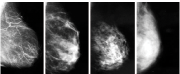
 Mammogram
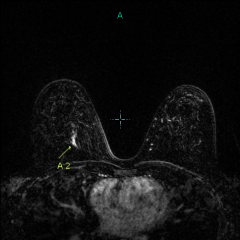 MRI
MRIThis 40-year-old woman has a strong family history of breast cancer. Her sister was diagnosed with breast cancer the previous year at age 44, and after learning more about breast cancer than she’d ever wanted to know, she advised our patient to start having yearly breast MRIs in addition to her mammogram because of her risk status (they also had family history in their maternal aunt and grandmother).
Our patient decided to have her first breast MRI, even though she’d had a negative mammogram the month before:

The arrow points to an abnormal 1.7cm area in her right breast that is “lighting up.”
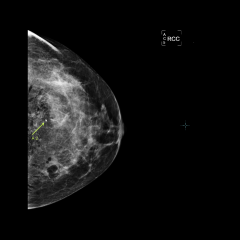
It could not be seen on her mammogram within her dense white breast tissue, even after knowing where it was located. See the mammogram pictures below, taken after her biopsy. The yellow arrow points to the metal clip that was placed at the site of the needle biopsy, which was performed with ultrasound guidance:


The diagnosis came back as High-grade Ductal Carcinoma in Situ (stage 0 breast cancer, i.e. very early cancer). The patient opted to have a lumpectomy and radiation therapy. She did not have to have chemotherapy, since the cancer was found so early. She plans to have a breast MRI every year in addition to her mammogram.